Home
Welcome to the Atomic-Scale Microscopy & Spectroscopy Group!
This group utilizes low-temperature scanning probe microscopy (SPM) that visualizes individual atoms and molecules in real space. We also perform localized spectroscopic measurements by irradiating the SPM junctions with laser light. This provides information on the sub-nanometer scale beyond the simple topography. The goal of this group is to understand the origin of characteristic chemical/physical phenomena at solid material surfaces and molecules adsorbed on the surfaces at the atomic level by high spatial resolution imaging and spectroscopy techniques.
News
Lab Exploration at Long Night of the Sciences
Jun 2025
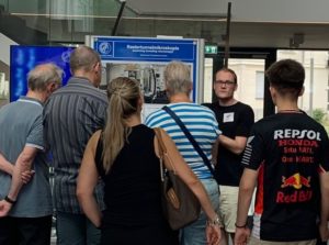
We conducted guided tours of our scanning tunneling microscopy laboratory for the general public at the Long Night of the Sciences in Berlin. Scientifically interested people of all ages could experience how atomically resolved images of a gold surface were recorded by the microscope in front of them in real time. This provided a unique opportunity to the audience where questions such as the working principles of the microscope and potential applications to novel materials science and quantum computing were discussed.
Publication: Elastic scattering-light detection meets noncontact atomic force microscopy
Jun 2025
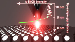
In a recent article published in Science Advances, we demonstrate scattering-type near-field optical microscopy (s-SNOM) based on low-temperature non-contact atomic force microscopy (nc-AFM). This resulted from a strong collaboration with Melanie Müller’s group in Department of Physical Chemistry and Electron Microscopy group in Department of Inorganic Chemistry. An ultralow tip-oscillation amplitude driven by a nc-AFM qPlus sensor allows for the sensitive detection of a plasmonic near-field localized in a narrow Ag-Ag gap. The nc-AFM-based s-SNOM achieves 1-nm resolution, surpassing the conventional s-SNOM resolutions.
Publication: Single-molecule-level vibrotational spectroscopy for hydrogen
May 2025

Our letter published in Physical Review Letters demonstrates the usefulness of tip-enhanced Raman spectroscopy (TERS) in characterizing physisorption systems. Weakly adsorbbed molecules, such as hydrogen on metal surfaces, are difficult to visualize even with scanning tunneling microscopy. Nevertheless, a plasmon-enhanced molecular spectroscopy technique called TERS well detects physisorbed hydrogen at the tip-sample junction. By analizing the vibrational/rotational spectra, we can also learn about the optical properties of the local tip-sample junction. Unlike previous TERS-measurement targets, the physisorption system exhibits poor chemical enhancement. Instead, the ultraconfined electromagnetic field at the atomic-scale local junction, namely, a plasmonic picocavity, dominates the Raman signal enhancement.
New group member: Hyeji Choi
May 2025

I am pleased to welcome Hyeji to our group as a doctral candidate. She works on single-molecule measurements using ultrahigh-vacuum low-temperature scanning probe microscopy. I wish her success!
Two posters from us brought prizes!
Oct 2024
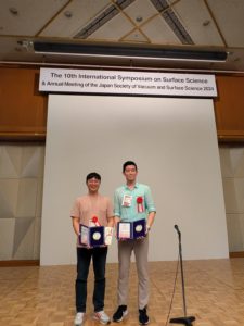
In an international conference, The 10th International Symposium on Surface Science (ISSS-10), Dr. Youngwook Park won Young Researcher Prize for his poster on the control of single-molecule photoswitching, and Mr. Kyungmin Kim, who visited us last year, also won Student Prize for his poster on tip-enhanced Raman spectroscopy measurements he conducted here. Their poster presentations were indeed remarkable. Congratulations, Youngwook and Kyungmin!