Equipment
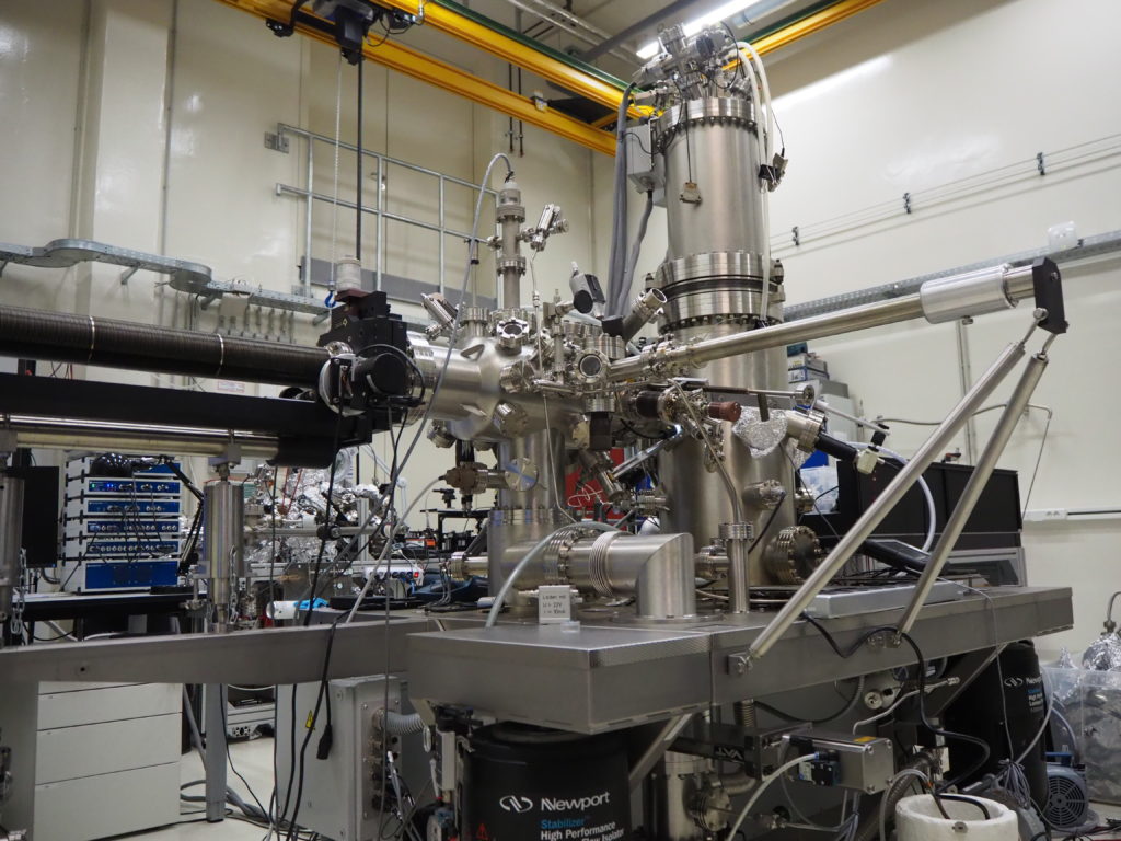
Our group conducts experimental research using ultra-high vacuum (UHV) chambers equipped with scanning probe microscopy (SPM). These machines are capable of scanning tunneling microscopy (STM) and non-contact atomic force microscopy (nc-AFM) measurements at low temperatures. They are also possible to irradiate laser light at the nanoscale junction between the probe tip and the sample, and to detect the light emitted from the junction. We are conducting the following experiments with these machines.
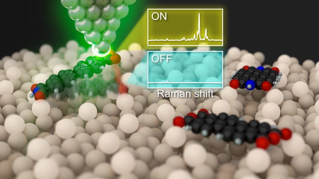
Sub-nanoscale vibrational spectroscopy
When silver or gold nanostructures are irradiated with visible light, surface plasmon polaritons are induced. By using the plasmons localized at the tip apex, the Raman signals of molecules in the vicinity of the tip apex can be enhanced. This technique is called tip-enhanced Raman spectroscopy (TERS), and is a powerful (sub-)nanoscale vibrational spectroscopy. With this method, we aim to obtain chemical information of solid surfaces and adsorbed molecules at the atomic scale, and understand the detection mechanism from the physical perspective. Furthermore, we develop new spectroscopic methods that can be applied to systems that were difficult to measure with conventional methods.
TERS of hydrogen molecules [see Reference for details]
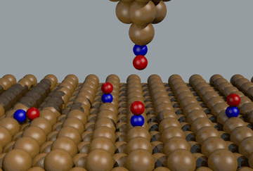
Control of chemical reactions and molecular switches
SPM is not limited to just observation; it can also induce chemical reactions and structural changes of molecules adsorbed on surfaces by applying local stimuli from the probe tip. This is an important clue for the fabrication of functional molecular devices, and would be an ultimate method to manipulate the structures and properties of individual molecules at the single-atom level. We aim to evaluate the response of individual molecules to local stimuli, in particular, external forces (mechanical stress) and near-field light.
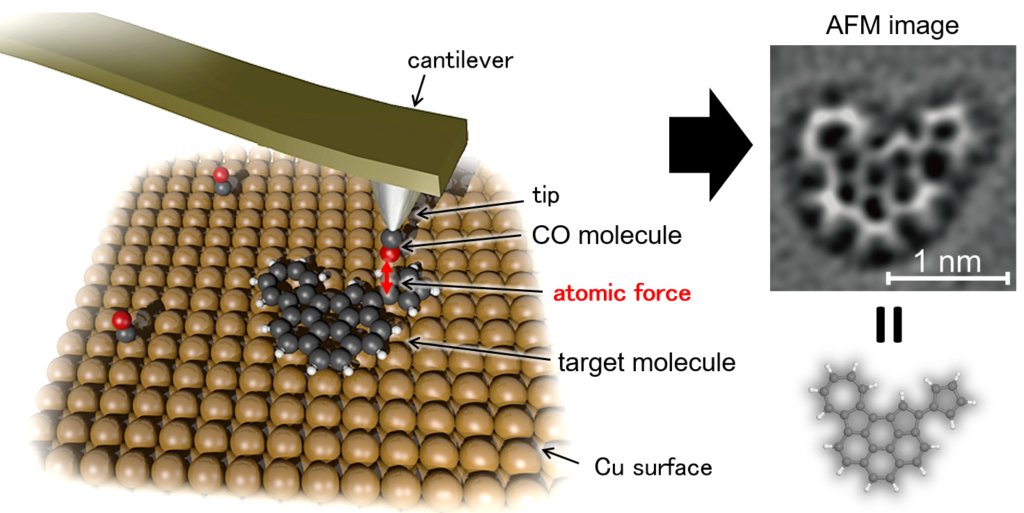
Ultrahigh spatial imaging
The direct observation with SPM can reveal the atomic arrangement of surfaces of solid materials and the atomic structure of molecules adsorbed on the surfaces. In particular, the carbon skeletons of aromatic hydrocarbon molecules can be visualized by precise detection of interatomic forces using nc-AFM. This technique can identify the molecular structures of reactants, intermediates, and products of chemical reactions occurring on the surface, including heterogeneous catalytic reactions.
We are working on extending the applicability of such a high-resolution imaging technique. As an example, we have shown that fragile molecular assemblies via relatively weak intermolecular interactions were imaged with unprecedented spatial resolution.