Home
Welcome to the Atomic-Scale Microscopy & Spectroscopy Group!
This group utilizes low-temperature scanning probe microscopy (SPM) that visualizes individual atoms and molecules in real space. We also perform localized spectroscopic measurements by irradiating the SPM junctions with laser light. This provides information on the sub-nanometer scale beyond the simple topography. The goal of this group is to understand the origin of characteristic chemical/physical phenomena at solid material surfaces and molecules adsorbed on the surfaces at the atomic level by high spatial resolution imaging and spectroscopy techniques.
News
Goodbye, Hyeji!
Apr 2026

Ms. Hyeji Choi had worked here on single-molecule mechanics measurements and obtained interesting results. Her experiences conducting the basic research experiments, attending a couple of workshops and conferences, and living in Berlin for one year must have been invaluable. We wish her all the best in her new role at the company from next month!
Two visiting students researching molecules
Oct 2025

We are currently conducting research with two visiting students. One is Mr. Hao Jiang, a PhD student from Shanghai University, who is investigating nanoscale photochemical reactions here for 6 months. The other is Mr. Naoki Hashimoto, a master’s student from Chiba University, who stays here for 2 months to conduct single-molecule spectroscopy of an organic compound he synthesized.
We hope that both make good progress with us!
We hope that both make good progress with us!
Publication: Overtone Raman spectroscopy for single molecules
Sep 2025

In Angewandte Chemie International Edition, our recent article was published as a “Hot Paper.” Overtone and combination-band transitions of molecules in Raman spectra are usually undetectable unless optimal resonance conditions are met. Since anharmonic motion depends on the potential energy surface, observing these modes helps elucidate energy transfer and chemical reaction processes. This study demonstrates that overtones and combination bands can be detected and analyzed with submolecular resolution using the point-contact tip-enhanced Raman spectroscopy.
Lab Exploration at Long Night of the Sciences
Jun 2025
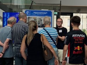
We conducted guided tours of our scanning tunneling microscopy laboratory for the general public at the Long Night of the Sciences in Berlin. Scientifically interested people of all ages could experience how atomically resolved images of a gold surface were recorded by the microscope in front of them in real time. This provided a unique opportunity to the audience where questions such as the working principles of the microscope and potential applications to novel materials science and quantum computing were discussed.
Publication: Elastic scattering-light detection meets noncontact atomic force microscopy
Jun 2025
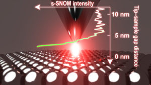
In a recent article published in Science Advances, we demonstrate scattering-type near-field optical microscopy (s-SNOM) based on low-temperature non-contact atomic force microscopy (nc-AFM). This resulted from a strong collaboration with Melanie Müller’s group in Department of Physical Chemistry and Electron Microscopy group in Department of Inorganic Chemistry. An ultralow tip-oscillation amplitude driven by a nc-AFM qPlus sensor allows for the sensitive detection of a plasmonic near-field localized in a narrow Ag-Ag gap. The nc-AFM-based s-SNOM achieves 1-nm resolution, surpassing the conventional s-SNOM resolutions.